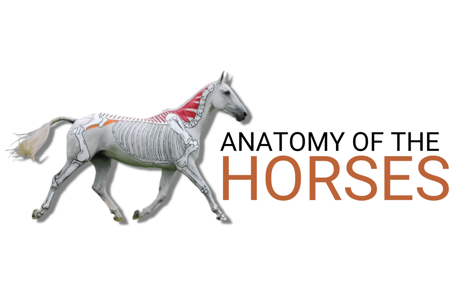
Anatomy of a horse
To analyze the morphology or the anatomy of the horse, it is important to distinguish the different parts of the animal. Indeed, knowing this animal well allows reducing the risk of exaggerated muscular efforts.
Anatomy of the horse: the 3 parts of the body
The horse’s body is divided into 3 parts:
- the forehand: head, neck, forelegs;
- the body: withers, back, belly;
- the hindquarters: croup, hip, thigh, patella, hock, cannon, fetlock, hoof, hind legs.
➤ Also Read: Can Horses Eat Cucumbers? The Surprising Truth About Feeding Cucumbers to Horses
The horse’s skeleton
On average, the horse grows until it is 5 years old, depending on the breed. In addition, there are about 200 bones in the horse’s skeleton that represent 8% of its weight. They are divided into 3 types:
- flat bones, such as those of the skull, pelvis or scapula;
- short bones, such as the vertebrae, the tarsus and the carpus;
- long bones, such as the femur, fibula and tibia.
The horse’s head
The horse’s head consists of the cranium, the face and the upper jaw made up of 34 bones. 2 mobile bones form the lower jaw.
In addition, its head has the following characteristics:
- To chew properly, the horse has well-developed jaws.
- It has well designed and powerful nostrils, knowing that its nasal cavities allow it to exchange large volumes of air.
- Its eyes are large and its ears, located on the back of the neck, are straight. It is the first two cervical vertebrae named atlas and axis that articulate the skull to the rachis.
The spine
The spine is composed of 54 bones. There are several vertebrae that make up the spine:
- 7 cervical: they help in the movement of flexion, extension, rotation and elevation of the neck;
- 18 dorsal vertebrae: the first 7 or 8 form the more or less prominent withers. The back is made up of 11 dorsals as well as the corresponding rib arches. Then come the loins which are generally muscular. These extend to the croup which goes to the tail. Finally, the tail is composed of 15 to 18 caudal vertebrae.
The horse has thirty six ribs. They work in pairs, 18 pairs. The first eight pairs are connected to the sternum while the other ten pairs come together to form the hypochondrium. This is what promotes good lung ventilation.
The forelegs
The forelegs start at the point of the shoulder while the forearm descends to the knee which, like the hock, must be large and prominent. Some bones, such as the ulna and fibula, are atrophied. Therefore, they are fused to the radius and the tibia. As for the front legs, they do not spread laterally because the horse does not have a clavicle. Finally, it is its powerful thorax that allows the horse to move more or less quickly.
Furthermore, the cannon, made up of hard and protruding tendons, goes down to the fetlock, separated from the foot by the pastern. The fetlock is composed of two large sesamoid bones that are connected to the pastern. Then comes the crown and finally the foot bone. Between the second and third phalanx, there is a small sesamoid bone, more commonly called the navicular bone.
The hind limbs
The hind limbs are composed of the hip bone, connected to the sacrum. This is what forms the croup. The coxal bone is itself made up of the ilium; the pubis and the ischium.
In addition, the coxo-femoral joint joins the coxal to the femur. We also distinguish the stifle which corresponds to the human knee. Covered by a patella, it is synchronized with the hock. Finally, the hock (tarsus) is composed of a series of joints maintained by ligaments. It is composed of 7 bones including the talus and the calcaneus.
Anatomy of the horse: muscle anatomy of a horse
The horse has 469 muscles that represent half of its weight. We distinguish 3 types of muscles which have a well determined role:
- the cardiac muscle;
- the white muscles;
- the red muscles.
The spinal column is equipped with tensor muscles: the ilio-spinal and the transverse spinous muscles.
The heart muscle
Located in the chest, the heart muscle is composed of striated fibers. This muscle functions by reflex with a heart rate of 30 to 40 beats per minute at rest and up to 220 beats per minute during very intensive work.
For a 500 kg horse, the heart can weigh between 3 and 5 kg.
The white muscles
The white muscles correspond to those of the viscera and control all organic functions. They ensure the internal functioning of the horse.
The red muscles
The red muscles (striated fibers) cause the movement by allowing the locomotion of the horse. They have different properties such as contractibility, elasticity, excitability and tonicity.
When the horse is in full effort and in movement, we can notice the muscles that work by ensuring 5 types of movements:
- extension, which opens a joint angle;
- flexion, which ensures the closing of the joint axis
- rotation, which allows for pivot effects, in the form of a circular movement;
- abduction, which ensures the spreading of the joint;
- adduction, which brings the joint closer together.
Finally, a distinction is made between muscles working in opposition and creating antagonistic movements, and congenital or agonistic muscles which are muscles acting in the same direction.
Horse head
The horse’s head is quite hefty; if the animal is large, it may weigh as much as 16 kg. This is something you should keep in mind, especially if you let the horse “hang” on a little bit and “carry” the weight of his head with your arms as you hold the reins the entire time. Such activity might leave you exhausted within an hour, which is why it’s so crucial to teach your horse to “carry” his own weight.
Depending on the animal’s race and kind, the head of a horse has different proportions to the rest of its body. Large heads enable cold-blooded horses to further ballast the front of their bodies, giving them tremendous pull strength. However, saddle horses with small heads compared to the rest of their bodies, like Arabs, are ideal for long or short distances (rides, races). The weight on the front of their bodies is lessened by their relatively small heads. However, it’s important to remember that a small head could indicate that the horse has anatomical flaws, including overbreeding and a short skeleton.
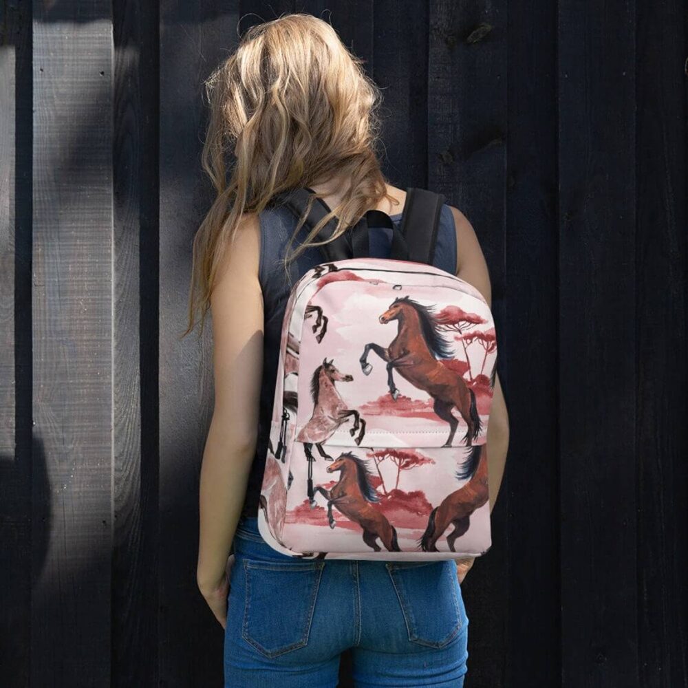
Carry your passion for horses wherever you go with our horse-themed accessories! Protect your device in style with our unique Horse Phone Cases or make a statement with a practical and stylish Horse Backpack. Start your day right with a favorite beverage in a Horse Mug or Cup, and keep your keys close with a charming Horse Keychain. For a touch of western flair, don’t miss our beautifully crafted Western Backpacks. Find the perfect accessory to match your equestrian spirit today!
Anatomy of horse head
The top (neurocranium) and bottom of the horse’s head can be separated (viscerocranium – the muzzle part).
The first section includes:
- The term “occiput” refers to the area behind the ears where the bridle (specifically, the occiput strap of the bridle) comes from. This area connects the horse’s head and neck.
- Between and in front of the ears lies the crown. It is a region whose lines are delineated by the eye fovea, the temporal fovea at the temples, the forehead, the eye sockets, and the eyes.
- On the front of the horse’s head, under the ears, is the forehead, which is often covered by a forelock that emerges from the crown.
The lower portion, or muzzle, consists of:
- Nose.
- Nostrils (along with the external part – nostril wings).
- the top and bottom lips (with chin).
- Branches of the jaw (the bottom borders of the jaw), the cheeks with the jaw below them, and the chin groove.
- Muzzle edges and groove.
Various horse head constructions
A horse’s head type is always influenced by his race. We distinguish 4 fundamental types:
Noble head, erect stance:
The forehead-nose line stays straight, making it the head shape that is the most precisely balanced. Horses of the breed and those used for racing typically have a long top and short bottom to their heads, along with a broad forehead.
Concave (dish) face:
Arabs and half-Arabs are distinguished by their broad foreheads, large eyes, and nostrils, but most notably by their concave noses and short falcate ears.
Roman nose:
The convex line of the nose, the short top part (neurocranium) and the long bottom part (viscerocranium), the narrow forehead, the small eyes and nostrils relative to the size of the head, and the long ears are characteristics of cold-blooded and racehorses like Lipizzaners and Kladrubers.
Pig head:
Its most distinguishing features are its very small nostrils and short muzzle.
Equine neck
His lever is the neck of the horse. Its muscles, length, and ratios to the rest of the horse’s body speak volumes about his health and propensity for sports, as well as the caliber of your training. A neck that is excessively short, for instance, could be problematic while jumping, where adequate basculing is essential. Basculing, which involves keeping the horse’s body stretched over an obstacle with its neck low and back arched, enables it to jump over extremely high hurdles while maintaining balance. While performing challenging dressage figures, a horse with a long neck may find it difficult to maintain balance.
Starting with the neck, you can relax the horse and improve the condition of his back. The neck is always essential for keeping one’s equilibrium, whether completing challenging dressage figures or jumping. You should alternate between stretching (relaxing) it, gradually shortening it (appropriate training), and finally extending it during training (stretching at the end of a ride).
Human horse neck anatomy
The following components make up the horse’s neck:
- Right and left sides,
- forelock/back of the neck,
- throat.
A horse that is ridden incorrectly must maintain a high head and neck, which prevents him from relaxing and causes him to develop exceptionally strong throat muscles.
Horse neck varieties
Three varieties of horse’s necks are distinguished:
- The horse should have an ideal (straight) neck because it indicates a straight trachea, which enables him to breathe in more air more quickly, which is essential during severe effort.
- Swan necks are frequently seen in Arab horses and are created when the top and bottom edges of the neck form an arch.
- Close-coupled neck: This condition leads the horse to carry his head high and trip frequently because the upper section of the neck is concave while the lower part of the neck is still convex. The horse has trouble breathing because the convex bottom section restricts airflow via the trachea.
Fortunately, swan and close-coupled necks are quite uncommon; the majority of horses have straight neck profiles. However, the settings are different:
- Highly set necks are neither a dressage or combined driving flaw.
- Neck positioned correctly.
- Low set necks are not considered a defect in Welsh ponies, horses used for difficult and long journeys, and carriage horses because they help the animals overcome resistance while pulling.
Anatomy of a horse hoof
An excellent illustration of Mother Nature’s technical prowess is the equine hoof. When you consider a horse’s size and weight in relation to the size of a hoof, as well as how quickly or far they can jump, you can see how much such little can hold. A horse’s capacity to survive and perform is significantly influenced by its hooves. Understanding hoof anatomy is crucial because a horse cannot exist without strong, healthy feet.
Even though the equine hoof appears simple, it is incredibly intricate. It consists of a number of different components that each serve a distinct function while also functioning symmetrically to maintain the horse’s soundness and health. For a better understanding of the hoof’s architecture and functions, it can be divided into three sections: the outside, the underside, and the interior.
External Buildings
Equine Wall
The hoof wall is the first aspect of the hoof that you notice. This is the tough, horny exterior that encases and safeguards the more fragile internal components, bears the weight of the horse, and cushions shock as it travels. The hoof wall is a continuously developing, keratinous substance that needs to be trimmed or naturally worn off; it lacks nerves and blood vessels. About 3/8 of an inch is added to a healthy hoof wall per month.
The hoof wall can be either white or black. Although it’s a common misconception, black hooves aren’t stronger than white ones. As farriers can attest, a horse’s four feet are all anatomically identical, hence the quality of a hoof is unaffected by its hue.
Due to its rigid nature, the hoof wall cannot enlarge when internal tissues are injured and swell. A horse may get lame if it injures the internal hoof structures. Rings or cracks should not be present in healthy hooves. Internal structures may become accessible to potentially harmful elements such as stones or viruses because of cracks. Rings on the hoof may be a sign that the horse has further health issues that are impacting his hooves, therefore you should visit your veterinarian.
Crown Band
The top of the hoof wall, where the hairline meets the hoof, is where the coronary band is located. This band, which encircles the top of the hoof wall, is often white. The hoof wall relies on it as its main source of nutrition and growth. It has a strong structural foundation and a lot of blood supply. The hoof wall may suffer damage through injury to the coronary band or from disruption of normal hoof growth, rendering the horse unrideable.
Periople
The periople serves as a protective covering for the hoof wall and the sensitive area directly below the coronary band. The periople helps give the freshly developed hoof wall tissue, which makes up the soft area, time to harden.
Laminar Layer or the inner wall
Hoofs have a flexible inner wall as opposed to a rigid outer wall. With a little more “give,” the inner wall can expand slightly with movement and absorb shock, safeguarding the sensitive inside areas of the hoof. The coffin bone is joined to the interior of the hoof wall by many laminae that resemble leaves, which are supported by the inner wall. Most of the weight of the horse is supported by these laminae.
Below the Hoof
Sole
The sole is the underside of the hoof, however because to its slight concavity, the majority of it does not make touch with the ground. The hoof wall’s structure and that of the sole are similar, but the hoof wall’s keratin is more easily scratched or worn away than the keratin in the sole. The sole is made to support internal weight transferred through the sole’s border rather than weight from the ground and aids in protecting the inner workings of the hoof.
The “white line,” which may really be somewhat yellow in hue, is a crucial part of the sole. The white line marks where the sole and hoof wall converge. The tissues in the white line region assist secure the sole to the inner wall of the hoof while also protecting the sole. Germs can infiltrate and separate the layers of the hoof wall when the white line area is damaged. Once this occurs, it may spread to other areas of the hoof and cause lameness in the horse.
Frog
The frog is readily seen when you take up the horse’s hoof; it is the hard, substantial, V-shaped structure pointing downward from the heels. It serves as a shock absorber as the horse moves, preserves the digital cushion beneath it, helps with traction and blood flow in the hoof. Your horse can feel the ground beneath his feet thanks to the delicate nerves in the frog that let him know where they are.
Middle Sulcus
The frog has grooves in the middle and on either side. The central sulcus and lateral sulci are located in the grooves down the middle and either side of the frog, respectively. The central sulcus should be shallow and rather wide. A narrow or deep sulcus, which can house germs and cause thrush, may be seen in horses with contracted hooves or sheared heels.
Bars
The bars are frog-parting extensions of the hoof wall that bend inward near the heel. The bars prevent the heels from overexpanding and tighten the heel region. Additionally, this region helps maintain the weight of the horse and contributes to the development of the hoof sole.
Internal Structure
Electronic Cushion
The region below the coffin bone near the back of the hoof is known as the digital cushion. It serves as one of the primary shock absorbers in the hoof and accomplishes precisely what its name suggests: it is a cushion of cartilaginous material with some “give.”
The digital cushion may be weakened in horses with long toes and low heels because the heels are carrying more weight than they should, which gradually reduces the cushion’s thickness. The digital cushion cannot regenerate once it has been “crushed.”
Death Bone
The bottom bone next to the toe that is enclosed in the hoof is known as the coffin (or “pedal”) bone. It helps to shape the hoof wall and is the biggest bone in the hoof. Special tissues that contribute to the laminae of the hoof wall and the tissues of the sole surround it. Lameness may come from anything that interferes with the coffin bone’s ability to function in conjunction with the hoof capsule, including significant changes in shoeing, sole punctures, and coffin bone rotation.
The little bone that is hidden below the coffin bone and the short pastern bone is termed the navicular bone, sometimes known as the distal sesamoid bone. The coffin bone is stabilized by the navicular bone, which also permits some tilting on unlevel terrain. The extensor tendon and the deep digital flexor tendon are the two main tendons that support and move the bones. The deep digital flexor tendon goes down the back of the leg and wraps around the navicular bone, bending and flexing the leg, as opposed to the extensor tendon, which attaches to the front of the coffin bone and straightens the leg.
The body language of the horse
The horse expresses himself a lot with his body. To decipher his emotions and desires, learn to recognize the basic gestures:
- His tail is up: he is in a good mood and in good health
- He has his tail between his buttocks: he is afraid
- Ears back: annoyed or afraid
- He puts his ears forward: he is attentive and interested
- He puts one ear forward, the other backward: he does not understand the orders which one gives him
- He is stamping his feet: he is impatient
- His eyes are bulging: he is very afraid
- His eyes are half closed: he is tired, sleepy or in poor health
Related Posts

Are Horses Aggressive? Spot Danger Signs in Their Body Language

Are Horses Smart Animals? New Studies Reveal Equine Intelligence

Where Can I Pet Horses Near Me: How to Find Local Farms & Rescues for a Visit

Where Horses Are Kept: From Luxury Stables to Open Pastures

Why Is Horse Sperm So Expensive: The Shocking Reality of Elite Stallion Breeding Fees

Why Is It Illegal to Bury a Horse? Environmental & Zoning Laws Explained

Where Are the Withers on a Horse: Why This Specific Point Is Crucial for Saddle Fit